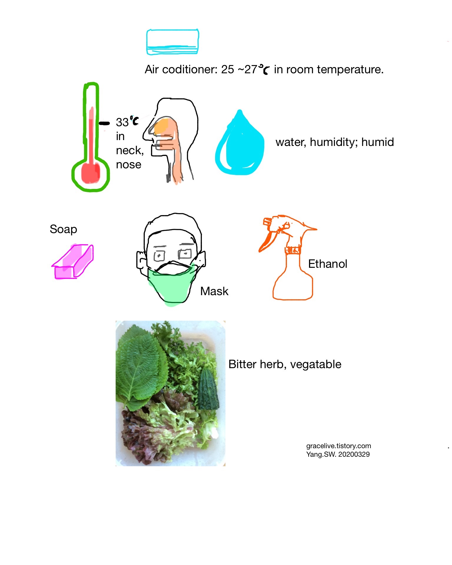
The optimal temperature for coronaviral proliferation is 33 °C. This temperature is the normal temperature in the upper respiratory tract. Hence, to keep the temperature in the upper airway more than 33°C is recommended.
Humidity
In cold and dry state, the viability of coronavirus is high.
Facial mask
Facial mask filters viral droplets and makes human upper airway keep warm and humid.
Air conditioner, Room temperature.
Room temperature is 20 ~ 22°C. In this temperature zone, the temperature of the upper airway is 33°C. To keep the upper airway in more than 33°C, the room temperature should be more than 23°C
As my personal experience considering both temperature and humidity, the adequate temperature of air-conditioner may range from 25 to 27°C.
Soap, Disinfectant
Ethanol, Disinfectant
Ethanol plays a role in breaking enevelope of coronavirus.
Bitter herbs, Food
Bitter herbs help our immunity boost.
Mediterranean diet
Meditteranean diet helps our immunity boost.
For detailed explanation, refer to category "2019-nCoV, COVID-19" within this blog.
Casanova et al demonstrated fecally contaminated liquid droplets of SARS cases are a potential vehicle for contagion besides respiratory droplets. This study reported that SARS coronaviruses remained infectious in water and sewage for days to weeks.[1]
For this reason, waterborne infection of COVID-19 coronaviruses should be checked meticulously to prevent further spread of COVID-19.
In addition to facial masks for blocking respiratory droplets, drinking water should be sterilized. At home, we must boil water or mix bitter herbs or lemons with water before drinking.
The government should pay attention to sterilize water supply source.
REFERENCE:
Casanova L, Rutala WA, Weber DJ, Sobsey MD. Survival of surrogate coronaviruses in water. Water Res. 2009 Apr;43(7):1893-8.
Besides the acute respiratory distress syndrome in the patients affected by COVID-19, the possibility of subclinical adrenal insufficiency should be considered. When patients are lethargic and dehydrated, intravenous glucose infusion can be considered. But if patients have adrenal insufficiency, intravenous glucose infusion can cause lethal damage, as the following reasons:
Based on the genetic and clinical similarity of COVID-19 to SARS coronaviruses, the autopsy findings in SARS affected patients are helpful to expect the things to come during the disease progression.
In the autopsy of SARS cases, the adrenal glands in patients revealed necrosis, infiltration of monocytes and lymphocytes loaded with SARS coronaviruses within vessels, and thrombosis in small veins.[1][2]
With this finding, we can infer the possibility of adrenal insufficiency.
In patients with adrenal insufficiency who have not received glucocorticoids, glucose infusion may cause high fever ("glucose fever") followed by collapse and death. Presumably, the glucose is metabolized, and the water dilutes the plasma, and the resultant osmotic gradient between the plasma and the cells causes the cells of the thermoregulatory centers in the hypothalamus to swell to such an extent that their function is disrupted.[3]
The author infers that the neuropathologic change of glucose fever may be similar to that of central pontine myelinolysis after too rapid medical correction of sodium deficiency (hyponatremia). Hyponatremia is often accompanied by adrenal insufficiency. Central pontine myelinolysis causes damage to myelin and neuron in the brainstem, especially pons and even extrapontine brain tissue.
On this point, intravenous fluid therapy in COVID-19 patients, subclinical hyponatremia conditions should be also considered.
The brain tissues infected with SARS coronaviruses are supposed to be susceptible to this hypothetical neural damage.
Gu J et al summarized many reports about observations of the central nervous system affected with SARS-coronaviruses, as follows:
RT-PCR has detected SARS-CoV genomic sequences in cerebral spinal fluid and in brain tissue specimens.The virus has been successfully isolated from brain tissue. Edema and focal degeneration of neurons have been observed in the brains of SARS autopsies. IHC(immunohistochemical stain),in situhybridization, and EM(electron microscopy) have confirmed viral infection of neurons. Gliocytes have also been found infected by SARS-coronaviruses.[4]
With the above reasons, the possibility of adrenal insufficiency and related neurologic damage can be applied to the cases with COVID-19.
For this reason, when patients with COVID-19 are lethargic and dehydrated, intravenous fluid therapy should be done without glucose except for overt hypoglycemic conditions and without rapid correction of hyponatremia.
To correct the adrenal insufficiency, the physiologic dose of steroids should be prescribed in the early phase of COVID-19. This may be also beneficial to prevent acute respiratory distress syndrome in COVID-19, for alveolar macrophages induce cytokine-related inflammatory responses that can be lessened by steroids.
P.S.
The physiologic dose of steroids the author recommends is methylprednisolone 1mg #2 (0.5mg intake two times) per day at a 60kg weighted person.
REFERENCE:
[1] Ding YQ, Wang HJ, Shen H, Li ZG, Geng J, Han HX, Cai JJ, Li X, Kang W, Weng DS, Lu YD, Wu DH, He L, Yao KT. The clinical pathology of severe acute respiratory syndrome (SARS): a report from China.J Pathol.2003;200:282–289.
[2]Gu J, Gong EC, Zhang B, Zheng J, Gao ZF, Zhong YF, Zou WZ, Zhan J, Wang SL, Xie ZG, Zhuang H, Wu BQ, Zhong HH, Shao HQ, Fang WG, Gao DX, Pei F, Li XW, He ZP, Xu DZ, Shi XY, Anderson VM, Leong ASY. Multiple organ infection and the pathogenesis of SARS. J Exp Med. 2005;202:415–424.
[4] Gu J, Korteweg C.Pathology and Pathogenesis of Severe Acute Respiratory Syndrome. Am J Pathol. 2007 Apr; 170(4): 1136–1147.
[3] Ganong WF. Chapter 20. The Adrenal Medulla & Adrenal Cortex, In Review of Medical Physiology, 22nd ed. Appleton & Lange, 2005, p 370.
Conventional coronavirus is known as an ss(single-stranded) RNA virus. Its incubation period is 2 ~ 5 days, whereas novel coronavirus is 4 ~ 14 days and SARS one is 2 ~ 7 days.
As supportive evidence of the incubation period of novel coronavirus longer than that of SARS coronavirus, there was a report three persons recovered from COVID-19 still had positive RT-PCR results for novel coronaviruses, even without symptoms and contagion into nearby family members.[1]
Its clinical implication has not been solved.
Despite novel coronaviruses are similar to SARS ones, why is the incubation period of novel coronaviruses longer than SARS ones?
What are the factors to explain the above phenomena?
In SARS, viral shedding is the nonlytic release of the vast majority of mature virions into the extracellular space via the Golgi apparatus from the ER.[2]. If novel coronavirus behaves in this way, the incubation period may be similar to that of SARS coronavirus.
If so, there must be the other pathway for viral shedding and replication to be continued except for the extracellular space, namely the intracellular space.
Which compartment of the intracellular space is probably suitable for viral shedding and replication?
The author supposes that the isolated hidden place for introverted viral shedding into the cell may be an intranuclear space, for the vast majority of viral release happens on the extracellular cytoplasmic border, as aforementioned.
The author infers the answer from the point that the main target cell organelle by novel coronavirus is endoplasmic-reticulum(ER).
Among four main proteins in coronaviruses, N protein forms a complex by binding to genomic RNA and M protein triggers the formation of interacting virions in this endoplasmic reticulum-Golgi apparatus intermediate compartment(ERGIC) with this complex.[3][4]
The characteristic electron microscopic findings of the alveolar epithelial cells in SARS cases are markedly dilated rough endoplasmic reticulum(RER) and smooth endoplasmic reticulum(SER).[5]
Uniquely, there was a comment on the membranous inclusion bodies in some nuclei.[5]
Considering the genetic & electron microscopic similarity of novel coronavirus to SARS coronavirus, the chance for nuclear inclusions to occur is high.
About the structure of nuclear inclusions, the reference journal did not include the related figure, and we can not clearly describe the electron microscopic feature to explain viral shedding into the intra-nuclear space.
Instead, the electron microscopic finding of nuclear inclusions in the cells may be an alternative tool to explain why nuclear inclusions in novel coronaviral infected cells can be a hidden reservoir for persistent viral replication.
About the detailed structure of nuclear inclusion, that in pituitary adenoma may be appropriate for a comparable explanation.
The nuclear inclusion in pituitary adenoma is composed of the ER-rich cytoplasm within the cell nucleus.
Yang et al(2003) analyzed the genesis of nuclear inclusion as the result of intracytoplasmic invagination into the nucleus. [6]
A part of the outline of the nuclear inclusion is composed of a double-layered membrane at the locus between the nucleus and the cytoplasm. Its origin can not be proved to be from whether ER or nuclear membrane.
The point is that nuclear inclusion includes ER-rich cytoplasmic components, which is continuous with the ER on the cytoplasmic side.
The author thinks that this nuclear inclusion with ER-rich cytoplasmic component can be a reservoir for persistent viral replication even during viral release into the extracellular space and cell division.
Despite the detrimental cytolytic cytotoxicity in COVID-19 cases, the presence of the long incubation period of novel coronaviruses can be explained by this hypothesis.
Hence, further research for novel coronaviruses should be performed to investigate the presence of nuclear inclusions in COVID-19 cases.
P.S.
If nuclear inclusions in COVID-19 cases will be found, please correspond to me about the next step for therapeutic strategy.
REFERENCE:
[1] Lan L, Xu D, Ye G, et al. Positive RT-PCR Test Results in Patients Recovered From COVID-19. JAMA. Published online February 27, 2020.
[2] Denison MR. CORONAVIRUS RESEARCH: KEYS TO DIAGNOSIS, TREATMENT, AND PREVENTION OF SARS. In: Institute of Medicine (US) Forum on Microbial Threats; Knobler S, Mahmoud A, Lemon S, et al., editors. Learning from SARS: Preparing for the Next Disease Outbreak: Workshop Summary. Washington (DC): National Academies Press (US); 2004.
[3] de Haan CA, Masters PS, Lili Kuo, Harry Vennema, Peter JM, Rottier. Coronavirus particle assembly: primary structure requirements of the membrane protein. J Virol. 1998; 72: 6838-6850.
[4] Escors D, Ortego J, Enjuanes L. The membrane M protein of the transmissible gastroenteritis coronavirus binds to the internal core through the carboxy-terminus. Adv Exp Med Biol. 2001; 494: 589-593.
[5] Ding Y, Wang H, Shen H, Li Z, Geng J, Han H, Cai J, Li X, Kang W, Weng D, Lu Y, Wu D, He L, Yao K. The clinical pathology of severe acute respiratory syndrome (SARS): a report from China. J Pathol. 2003 Jul;200(3):282-9.(PDF free download)
[6] Yang SW, Yang KM, Kang Hy, Kim TS. Intranuclear cytoplasmic pseudoinclusions in pituitary adenomas. Yonsei Med J. 2003 Oct 30;44(5):816-20.(PDF free download)
te Velthuis et al reported that Zn(2+) inhibits the replication of SARS coronaviruses in cell culture study. They found that the SARS-CoV RdRp elongation was inhibited and template binding reduced by Zn(2+).[1]
Mocchegiani et al found that old people aged 60-65 years with specific IL-6 polymorphism (GG allele carriers; named C-) had a low level of zinc and the proinflammatory status of IL-6. With zinc supplementation, the inflammatory responses by IL-6 were ameliorated into the healthy condition.[2]
Old people have a tendency to have an increased level of IL-6 associated with the aging process.[3]
It has known that increased IL-6 production by Th2 cells and macrophages.[4]
This Th2 shifting immunity is susceptible to viral infection.
About the immunity of COVID-19, Xu et al revealed Th2 immune skewness polarized by IL-6 and TGF-β, which is vulnerable to viral infection.[5]
With a constellation of the above data, although there is no report about the relationship between zinc and novel coronaviral infection(COVID-19, Wuhan Coronaviral infection), the author thinks that zinc can be a supportive nutritional mineral for anti-viral immunity in COVID-19.
REFERENCE:
[1] te Velthuis AJ, van den Worm SH, Sims AC, Baric RS, Snijder EJ, van Hemert MJ. Zn(2+) inhibits coronavirus and arterivirus RNA polymerase activity in vitro and zinc ionophores block the replication of these viruses in cell culture. PLoS Pathog. 2010 Nov 4;6(11):e1001176.
[2] Mocchegiani E, Romeo J, Malavolta M, Costarelli L, Giacconi R, Diaz LE, Marcos A. Zinc: dietary intake and impact of supplementation on immune function in elderly. Age (Dordr). 2013 Jun;35(3):839-60. doi: 10.1007/s11357-011-9377-3. Epub 2012 Jan 6. PMID: 22222917; PMCID: PMC3636409.
[3] Franceschi C. Inflammaging as a major characteristic of old people: can it be prevented or cured? Nutr Rev. 2007 Dec;65(12 Pt 2):S173-6.
[4] Mocchegiani E, Muzzioli M, Cipriano C, Giacconi R. Zinc, T-cell pathways, aging: role of metallothioneins. Mech Ageing Dev. 1998;106:183–204.
[5] Xu Z, Shi L, Wang Y, Zhang J, et al. Pathological findings of COVID-19 associated with acute respiratory distress syndrome. Lancet Respir Med. 2020;S2213-2600(20)30076-X.
In the pulmonary cases of COVID-19, Xu et al found the cytotoxic T cells with cytolytic activity which is associated with Th17 CD4 T cellspolarized by IL-6 and TGF-β. This immunopathology led to diffuse alveolar and interstitial damage with pulmonary edema, pathologically and subsequent acute respiratory distress syndrome(ARDS) and secondary pneumonia, clinically.[1]
As a nutritional candidate to modulate both IL-6 and TGF-β, vitamin D can be considered.
Dalvi et al reported that the serum level of IL-6 was significantly increased and vitamin D3 decreased in tuberculosis multidrug-resistant group. Conversely, we can expect that the elevated serum level of vitamin D may reduce that of IL-6.[2]
Hu et al mentioned the protecting role of vitamin D in diabetic nephropathy. One of the mechanisms to suppress pro-inflammatory responses was to inhibit production of TGF-β.[3]
In conclusion, because the problematic immunopathology in the lung injury by COVID-19 is the elevated serum levels of IL-6 andTGF-β, we can infer that vitamin D to counteract these inflammatory responses may play a role somewhat in ameliorating the lung injury by COVID-19. About this, further studies will be necessary.
REFERENCE:
[1] Xu Z, Shi L, Wang Y, Zhang J, et al. Pathological findings of COVID-19 associated with acute respiratory distress syndrome. Lancet Respir Med. 2020;S2213-2600(20)30076-X.
[2] Dalvi SM, Ramraje NN, Patil VW, Hegde R, Yeram N. Study of IL-6 and vitamin D3 in patients of pulmonary tuberculosis. Indian J Tuberc. 2019 Jul;66(3):337-345.
[3] Hu X, Liu W, Yan Y, Liu H, Huang Q, Xiao Y, Gong Z, Du J. Vitamin D protects against diabetic nephropathy: Evidence-based effectiveness and mechanism. Eur J Pharmacol. 2019 Feb 15;845:91-98.
About the usefulness of megadose vitamin C in common colds or flu-like symptoms, there have been many debates.
With the high potency of the antioxidant effect in vitamin C, vitamin C seems to be a healthful nutrient to suppress the inflammatory reaction in cancer or infection, including viral respiratory diseases.
Gorton & Jarvis reported that vitamin C use in the early phase of virus-induced respiratory infections can be helpful to prevent further disease progression or to relieve symptoms of common colds or flu-like symptoms.[1]
In contrast, Douglas et al analyzed that vitamin C plays some role in respiratory defense mechanisms, but vitamin C at the onset of common colds did not show clinically significant benefit.[2]
In summary, the beneficial role of vitamin C in common colds or virus-induced respiratory infections has not been clearly established.
But Oudemans-van Straaten et al commented that vitamin C can restore vascular responsiveness to vasoconstrictors, preserve endothelial barrier by maintaining cyclic guanylate phosphatase and occludin phosphorylation and preventing apoptosis. Furthermore, they mentioned the role of high-dose vitamin C in augmenting antibacterial defense. With this context, they argued that vitamin C can be applied for ischemia/reperfusion injury and sepsis.[3]
Although there are many conflicting reports on the beneficial role of vitamin C in common colds, at least vitamin C can be used as an antioxidant to maintain vascular integrity and plausibly to prevent aggravation of secondary bacterial infection.
REFERENCE:
1. Gorton HC, Jarvis K. The effectiveness of vitamin C in preventing and relieving the symptoms of virus-induced respiratory infections. J Manipulative Physiol Ther. 1999 Oct;22(8):530-3.
2. Douglas RM, Hemila H, D'Souza R, Chalker EB, Treacy B. Vitamin C for preventing and treating the common cold. Cochrane Database Syst Rev. 2004 Oct 18;(4):CD000980.
3. Oudemans-van Straaten HM, Spoelstra-de Man AM, de Waard MC. Vitamin C revisited. Crit Care. 2014 Aug 6;18(4):460.
Chapter 12 of Exodus in Bible reports on Passover day. Here God ordered the Israelite to avoid disaster on Passover day.
Chapter 12: verse 12th and 13th of Exodus, Bible says, "12on that same night I will pass through Egypt and strike down every firstborn - both men and animals - and I will bring judgment on all the gods of Egypt. I am the LORD.13The blood will be a sign for you on the houses where you are; and when I see the blood, I will pass over you. No destructive plague will touch you when I strike Egypt" (Figure 1).
The Passover means the disaster skipped the Israelite who kept the orders of God.
God orders the Israelite to paint the blood of lamb or goat on the sides and tops of the doorframe.
Why did God order this?
I interpreted this question medically. In terms of pathology, my opinion on these two problems is as follows;
Blood color varies from red to dark purple according to oxygen saturation. The main component of blood hue is caused by RBC (Red Blood Cell). When oxygen is rich in blood, blood color is red by binding O2to iron in hemoglobin. Conversely, when oxygen is deficient, blood hue is dark purple. When we have cut wound in the skin, the blood coagulates slightly sticky. This occurs mainly by the fibrin component. The fibrin particles are interlocked and form chains of fibrin, a structure of meshwork (Fig.2.). This meshwork structure filters blood cells, especially platelets and RBCs. All this process leads to blood coagulation. The characteristics of the fibrin component endow the blood glutinous.
Sticking nature of blood by fibrin and blood other components, namely viscous force, makes blood to acquire the filtering power to capture fine dust of air including microorganisms (bacteria and virus particles).
This may dilute the density of microbial pathogen particles.
As a principle of vaccine, the diluted amount of microbial pathogens may work as an immune-boosting effect rather than causing disease.
In this situation, I infer the people who did not paint blood on the doorframe may inhale contaminated air and be exposed to the infectious state. In contrast, people, namely the Israelites who paint blood on the doorframe, may inhale the air of diluted infectious microbial particles, which worked as a vaccine effect.
Furthermore, God designates that doorframe painting with blood should be done at night (Exodus, chapter 12; verse 6th:Take care of them until the fourteenth day of the month, when all the people of the community of Israel must slaughter them at twilight). To keep the viscosity of blood, the blood should preserve the liquidness. The liquid state may most be maintained at the cool state. The cool state in a day may be kept at the night time after sunset.
For the above reason, I infer God ordered blood paint on the doorframe at night in a day.
Copyright (C-2013-017025)PASSOVER DAY – Medical Interpretation by a Pathologist – (English)
Facial Mask in the year 2020:
As of March. 2020, novel coronavirus infection is pandemic. As a primary preventive method, the importance of the facial mask is raised. The author thinks that the facial mask may play a role in decreasing the number of viral droplets in inflowing air like the doorframe painted with blood in Passover.